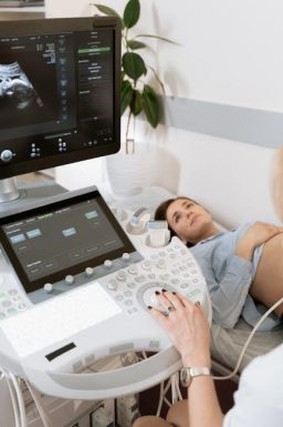
Ultrasound-guided BBL represents a significant advancement, offering surgeons enhanced precision and safety during fat transfer procedures.
Recent online resources, including Rutube videos from 2021, 2023, and 2024, demonstrate a growing interest in innovative surgical techniques.
This method utilizes real-time imaging to optimize fat placement, improving aesthetic outcomes and minimizing potential complications, as highlighted by various online guides.
What is a Brazilian Butt Lift (BBL)?
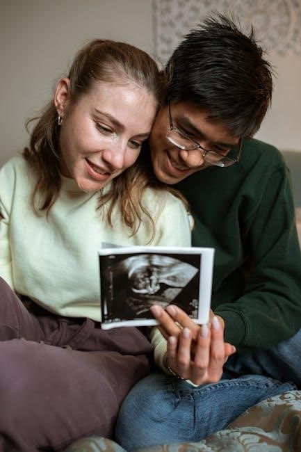
A Brazilian Butt Lift (BBL) is a cosmetic surgical procedure designed to enhance the shape and size of the buttocks using a patient’s own fat. This involves liposuction to harvest fat from areas like the abdomen, flanks, or thighs, followed by careful injection into the buttocks;
The goal is to create a more proportionate and aesthetically pleasing figure. While traditionally performed by anatomical landmarks, the integration of ultrasound guidance represents a modern evolution. Online resources emphasize the importance of precision in fat placement for optimal results, a key benefit offered by ultrasound technology.
This technique aims to improve graft survival and reduce the risk of complications.
The Role of Ultrasound in BBL Procedures
Ultrasound technology plays a crucial role in modernizing BBL procedures by providing real-time visualization of underlying anatomical structures. This allows surgeons to precisely target fat injection sites, avoiding blood vessels and ensuring optimal graft placement.
Unlike traditional methods relying on palpation, ultrasound offers a dynamic view, enhancing safety and accuracy. Recent online discussions highlight its utility in mapping vascular structures, minimizing the risk of complications.
Furthermore, ultrasound aids in assessing fat distribution and viability, contributing to improved aesthetic outcomes and patient satisfaction.
Pre-Operative Assessment & Planning
Detailed pre-operative planning, utilizing ultrasound imaging, is essential for successful ultrasound-guided BBLs. This includes careful patient selection and thorough evaluation of fat grafting sites.
Patient Selection Criteria for Ultrasound-Guided BBL
Ideal candidates for ultrasound-guided BBL possess good overall health and realistic expectations. A comprehensive medical history review is crucial, excluding individuals with certain conditions.
Patients should have sufficient donor fat available for harvesting, assessed through physical examination and potentially preliminary ultrasound imaging.
Those with significant vascular issues or bleeding disorders may not be suitable due to increased risks.
Furthermore, a stable weight is preferred, as fluctuations can affect long-term results. Careful consideration of skin elasticity and body composition is also vital for optimal outcomes.
Ultrasound Imaging for Fat Grafting Site Evaluation
Pre-operative ultrasound meticulously maps the recipient sites, identifying critical vascular structures and anatomical landmarks. This detailed assessment minimizes the risk of intravascular fat injection, a serious complication.
Ultrasound reveals the depth of subcutaneous tissue and muscle layers, guiding precise needle placement during fat grafting. It also helps visualize existing fat compartments and identify areas needing augmentation.
The technique allows surgeons to detect potential anatomical variations and avoid areas with compromised blood supply, ensuring optimal graft survival and aesthetic results.
3D/4D Ultrasound for Volume Estimation
Advanced ultrasound technologies, specifically 3D and 4D imaging, provide surgeons with accurate estimations of buttock volume deficits. This precise quantification is crucial for determining the necessary amount of harvested fat for optimal results.
These modalities create detailed three-dimensional reconstructions of the gluteal region, allowing for personalized treatment planning. Surgeons can visualize the potential outcome and adjust the grafting strategy accordingly.
Dynamic 4D imaging assesses tissue movement and elasticity, further refining volume estimations and ensuring a natural-looking augmentation.
Surgical Technique: Ultrasound Guidance
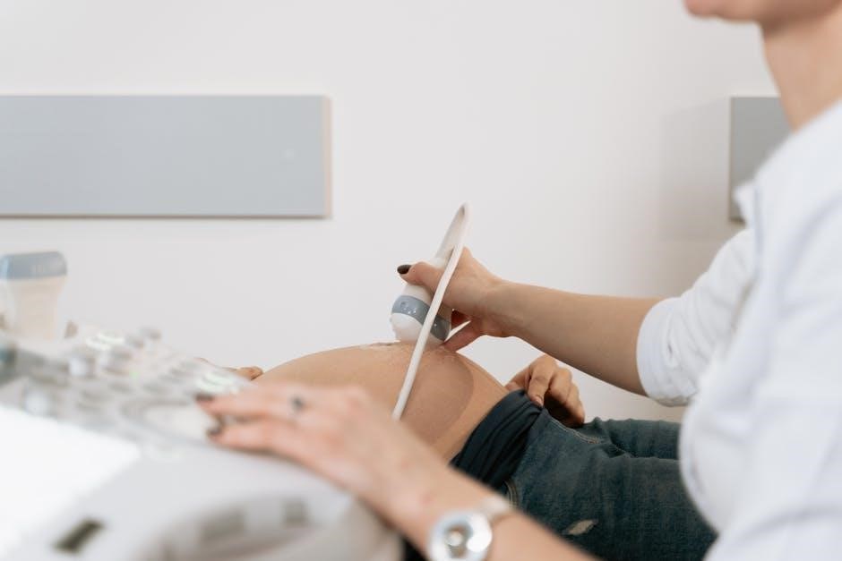
Ultrasound guidance revolutionizes BBL surgery, enabling real-time visualization of fat harvesting and precise, layered grafting. This technique enhances safety and aesthetic precision.
Surgeons utilize ultrasound to monitor fat distribution, ensuring optimal placement and minimizing complications.
Real-Time Ultrasound Visualization During Fat Harvesting
Real-time ultrasound dramatically improves the safety and efficiency of fat harvesting in BBL procedures. Utilizing high-frequency sound waves, surgeons can visualize the subcutaneous fat layers with exceptional clarity.
This allows for precise cannula placement, avoiding critical structures like blood vessels and nerves. The dynamic imaging provided by ultrasound ensures surgeons target optimal fat reserves, minimizing trauma to surrounding tissues.
Furthermore, it aids in assessing fat quality and quantity during the harvesting process, contributing to more predictable and aesthetically pleasing results. This technique represents a significant advancement over traditional, blind fat harvesting methods.
Precise Fat Grafting with Ultrasound Control
Ultrasound control revolutionizes fat grafting precision during BBL, enabling surgeons to visualize the deposition of fat in real-time. This allows for targeted injections into specific muscle layers and subcutaneous planes, optimizing volume and contour.
The technology minimizes the risk of injecting fat into unwanted areas, such as blood vessels, significantly enhancing patient safety. Surgeons can confirm proper graft placement and distribution, achieving symmetrical and natural-looking results.
This level of control is crucial for sculpting the buttocks and achieving the desired aesthetic outcome, surpassing the accuracy of traditional techniques.
Layered Fat Injection Guided by Ultrasound
Ultrasound guidance facilitates a layered approach to fat injection, mimicking the natural anatomy of the buttocks. Surgeons can precisely deposit fat into the superficial, middle, and deep compartments, creating a stable and aesthetically pleasing shape.
This technique ensures optimal graft survival by placing fat in areas with sufficient blood supply; Visualizing these layers in real-time minimizes the risk of unevenness or distortion.
The layered injection method, guided by ultrasound, promotes a more natural contour and long-lasting results, enhancing patient satisfaction.
Ultrasound Monitoring of Fat Distribution
Real-time ultrasound allows surgeons to meticulously monitor fat distribution during the BBL procedure. This dynamic visualization ensures even graft placement and prevents clumping or migration, crucial for achieving symmetrical results.
By observing the fat’s interaction with surrounding tissues, surgeons can adjust injection techniques as needed, optimizing volume and contour. This continuous assessment minimizes the risk of asymmetry and enhances the overall aesthetic outcome.
Precise monitoring contributes to improved graft survival and a more natural-looking result.

Benefits of Ultrasound Guidance
Ultrasound guidance in BBL significantly improves graft survival rates, reduces vascular risks, and enhances aesthetic symmetry. It offers a minimally invasive approach for optimal results.
Improved Graft Survival Rates
Ultrasound guidance demonstrably boosts fat graft survival by allowing surgeons to meticulously assess recipient site vascularity in real-time. This precise visualization ensures optimal blood supply to the transferred fat cells, fostering their integration into the native tissue.
Traditional BBL techniques rely on tactile feel and visual estimation, which can be less accurate. Ultrasound enables surgeons to identify and avoid areas with insufficient blood flow, minimizing fat necrosis and maximizing the volume of successfully engrafted fat. This leads to more predictable and long-lasting results, enhancing patient satisfaction.
Furthermore, careful monitoring of fat distribution with ultrasound helps prevent overcorrection or undercorrection, contributing to a more natural and balanced aesthetic outcome.
Reduced Risk of Vascular Complications
Ultrasound guidance significantly mitigates the risk of intravascular fat injection, a potentially life-threatening complication associated with traditional BBL procedures. Real-time visualization of blood vessels allows surgeons to precisely navigate the cannula, avoiding direct puncture of veins and arteries.
Doppler ultrasound, a key component of this technique, maps vascular structures, providing a clear roadmap for safe fat deposition. This is crucial in areas with dense vascular networks, such as the gluteal region.
By confirming cannula placement outside of vessels before injecting, ultrasound dramatically enhances patient safety and reduces the incidence of serious adverse events.
Enhanced Symmetry and Aesthetic Outcomes
Ultrasound guidance allows for meticulous fat distribution, leading to improved symmetry and a more natural-looking result in BBL procedures; Surgeons can visualize fat placement in real-time, ensuring even volume distribution across both buttocks.
Precise layering of fat grafts, guided by ultrasound imaging, creates optimal contouring and projection. This technique addresses asymmetries that may be present due to anatomical variations or prior fat grafting.
The ability to monitor fat distribution intraoperatively ensures a balanced and aesthetically pleasing outcome, maximizing patient satisfaction.
Minimally Invasive Approach
Ultrasound-guided BBL contributes to a less invasive surgical experience compared to traditional techniques. The precise visualization offered by ultrasound reduces the need for large incisions and extensive tissue dissection during fat harvesting and grafting.
Targeted fat placement minimizes trauma to surrounding tissues, resulting in less post-operative pain, swelling, and bruising. This approach allows for quicker recovery times and a return to normal activities.
The enhanced precision also reduces the risk of complications, further supporting a minimally invasive philosophy in BBL surgery.
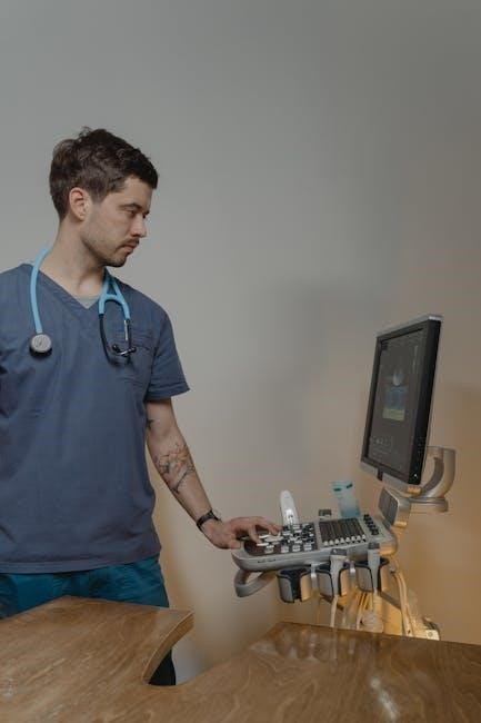
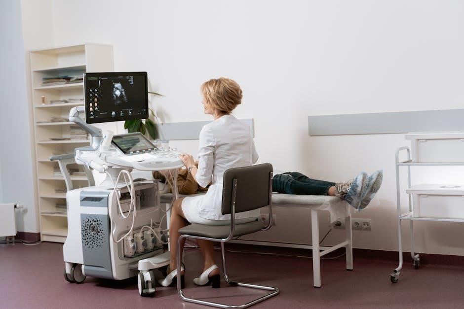
Ultrasound Technology & Equipment
Ultrasound-guided BBL relies on specialized machines and transducers for real-time visualization. Doppler ultrasound is crucial for vascular mapping, ensuring safe fat grafting procedures.
Modern systems offer advanced imaging capabilities, enhancing precision and patient safety during the entire process.
Types of Ultrasound Machines Used in BBL
Ultrasound-guided BBL procedures utilize a range of ultrasound technologies, primarily focusing on high-resolution imaging. Linear array transducers, offering superior superficial resolution, are frequently employed for visualizing subcutaneous fat layers and vascular structures during both fat harvesting and grafting phases.
Convex array transducers provide a wider field of view, beneficial for initial assessment and deeper tissue evaluation. Portable ultrasound systems are gaining popularity due to their flexibility and ease of integration into the operating room workflow. Furthermore, machines with Doppler capabilities are essential for identifying and avoiding critical blood vessels, minimizing the risk of vascular complications.
Advanced systems may also incorporate 3D/4D imaging for enhanced volume estimation and precise fat placement.
Ultrasound Transducers and Their Applications
Ultrasound transducers are pivotal in ultrasound-guided BBL, converting electrical energy into sound waves and receiving returning echoes to create images; High-frequency linear transducers (typically 7-12 MHz) excel at visualizing superficial structures like fat and vessels during harvesting.
Low-frequency curved transducers (2-5 MHz) offer deeper penetration for assessing muscle layers and overall anatomical context. Doppler transducers assess blood flow, crucial for avoiding vascular injury during cannulation and injection. The choice depends on the anatomical location and desired imaging depth.
Proper transducer selection ensures optimal image quality and procedural safety.
Doppler Ultrasound for Vascular Mapping
Doppler ultrasound is integral to ultrasound-guided BBL, providing real-time visualization of blood flow within tissues. This technique meticulously maps vascular structures before and during fat harvesting and injection, significantly reducing the risk of intravascular fat embolism – a serious complication.
Color Doppler displays blood flow direction and velocity, while Power Doppler enhances sensitivity for detecting subtle vessels. Surgeons utilize this to avoid perforating arteries and veins during cannula insertion, ensuring patient safety.
Precise vascular mapping optimizes graft survival and minimizes post-operative complications.
Potential Risks and Complications
Despite advancements, ultrasound-guided BBL carries risks like vascular complications, seroma, hematoma, and fat embolism. Vigilant ultrasound monitoring aids early detection and management.
Proactive strategies minimize these risks, ensuring patient safety throughout the procedure and recovery phases.
Addressing Vascular Complications with Ultrasound
Real-time ultrasound visualization is paramount in mitigating the most serious risk associated with BBL – intravascular fat embolism. Doppler ultrasound allows surgeons to meticulously map vascular structures before and during fat grafting.
This precise vascular mapping enables avoidance of direct cannulation into blood vessels during fat injection. Should inadvertent intravascular injection occur, ultrasound can immediately identify it, prompting immediate cessation of injection and supportive care. Early detection significantly improves patient outcomes.
Continuous monitoring throughout the procedure, coupled with a thorough understanding of ultrasound principles, is crucial for safe and effective BBL surgery.
Seroma and Hematoma Detection via Ultrasound
Post-operative ultrasound is a highly effective tool for early detection of seromas and hematomas, common complications following BBL procedures. Ultrasound imaging differentiates between fluid collections (seromas) and blood accumulations (hematomas) based on their echogenicity – how they reflect sound waves.
Early identification allows for timely intervention, such as aspiration, preventing prolonged discomfort, infection risk, and potential graft compromise. Ultrasound’s non-invasive nature makes it ideal for repeated assessments.
Regular ultrasound scans in the post-operative period contribute to improved patient recovery and minimized complications.
Fat Embolism Prevention Strategies
Ultrasound guidance plays a crucial role in minimizing the risk of fat embolism during BBL. Precise fat harvesting and gentle injection techniques, facilitated by real-time visualization, reduce the likelihood of fat entering the venous system.
Doppler ultrasound is utilized to meticulously map vasculature, avoiding cannulation of large veins during fat grafting. Careful aspiration and avoiding excessive pressure during injection are paramount.
Strict adherence to established surgical protocols, combined with ultrasound’s enhanced precision, significantly contributes to patient safety and reduces this rare but serious complication.

Post-Operative Care & Monitoring
Post-operative ultrasound assesses graft viability and detects complications like seroma or hematoma. Monitoring ensures optimal healing and addresses any concerns promptly, improving outcomes.
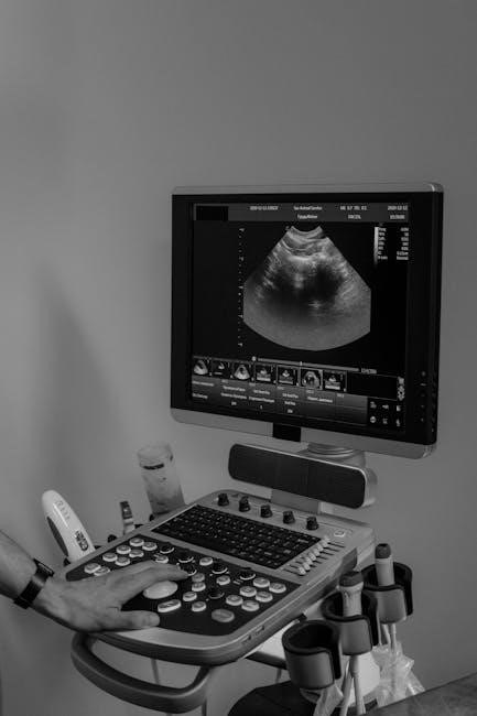
Ultrasound Evaluation of Graft Viability Post-Op
Post-operative ultrasound imaging plays a crucial role in evaluating the success of an ultrasound-guided BBL. It allows surgeons to non-invasively assess the fat grafts, determining blood flow and viability within the newly transferred tissue. Doppler ultrasound specifically maps vascularity, confirming adequate perfusion to the grafted fat cells.
Early detection of compromised grafts is vital; ultrasound can identify areas with reduced blood supply, potentially requiring additional supportive measures. This proactive approach minimizes fat absorption and maximizes long-term volume retention. Regular follow-up scans, typically scheduled within the first week and then at intervals thereafter, provide valuable data on graft health and overall healing progress.
Monitoring for Post-Operative Complications
Post-operative ultrasound is invaluable for detecting potential complications following an ultrasound-guided BBL. It efficiently identifies seromas – fluid collections – and hematomas – blood accumulations – which can occur in the grafted areas. Early detection allows for timely intervention, such as aspiration, minimizing discomfort and promoting healing.
Ultrasound also aids in monitoring for vascular issues, though less common with guided techniques. Furthermore, it can assess the compression garment’s effectiveness, ensuring adequate support and minimizing swelling. Regular scans provide peace of mind, allowing surgeons to proactively address any concerns and optimize patient recovery.
Compression Garment Use and Ultrasound Findings
Ultrasound assessment plays a crucial role in evaluating the effectiveness of compression garment use post-BBL. Imaging reveals how well the garment minimizes swelling and supports the newly grafted fat. Findings can indicate areas requiring adjusted compression or potential fluid accumulation despite garment adherence.
Ultrasound can detect subtle irregularities in fat distribution influenced by garment pressure. This allows for personalized recommendations, optimizing garment wear and promoting graft survival. Consistent monitoring ensures the garment contributes positively to the healing process and desired aesthetic outcome.
Advanced Ultrasound Techniques
Cutting-edge techniques like Contrast-Enhanced Ultrasound (CEUS) and Elastography refine ultrasound-guided BBL. These modalities offer detailed graft assessment,
enhancing visualization of vascularity and tissue consistency for optimal results.
Contrast-Enhanced Ultrasound (CEUS) in BBL
CEUS utilizes microbubble contrast agents injected intravenously to amplify ultrasound signals, providing a dynamic assessment of blood flow within grafted fat tissues. This technique significantly improves visualization of perfusion, allowing surgeons to evaluate graft viability in real-time.
Unlike traditional Doppler ultrasound, CEUS offers superior sensitivity in detecting subtle changes in microcirculation, crucial for identifying areas of compromised graft survival. It helps differentiate between functioning and non-functioning fat, guiding further adjustments during the procedure. CEUS is a valuable tool for optimizing graft take and enhancing long-term aesthetic outcomes in BBL procedures.
Elastography for Assessing Graft Consistency
Elastography is an advanced ultrasound technique that maps tissue stiffness, providing valuable insights into the structural integrity of grafted fat. By measuring tissue elasticity, surgeons can differentiate between viable, well-integrated fat and areas of fibrosis or necrosis.
This non-invasive method helps assess graft quality and predict long-term stability, contributing to more predictable and durable aesthetic results. Elastography complements traditional ultrasound imaging, offering a comprehensive evaluation of the BBL outcome and aiding in post-operative monitoring of graft health.
Fusion Imaging (Ultrasound & CT/MRI)
Fusion imaging combines the real-time capabilities of ultrasound with the detailed anatomical information from CT or MRI scans. This innovative approach creates a comprehensive 3D visualization of the patient’s anatomy and the grafted fat distribution.
By overlaying ultrasound images onto pre-operative CT/MRI data, surgeons can precisely target fat injections, avoid critical vascular structures, and optimize graft placement. This technique enhances accuracy, minimizes risks, and improves the overall aesthetic outcome of the ultrasound-guided BBL procedure.

Comparing Ultrasound-Guided BBL to Traditional BBL
Ultrasound-guided BBL demonstrates superior accuracy and precision compared to traditional methods, potentially leading to faster recovery times and improved aesthetic results.
Enhanced visualization minimizes complications, offering a safer and more predictable outcome for patients.
Accuracy and Precision Differences
Traditional BBL relies heavily on the surgeon’s tactile feel and visual assessment, introducing a degree of subjectivity. Ultrasound-guided BBL, however, offers real-time visualization of fat graft placement, significantly enhancing precision.
This allows surgeons to accurately target specific muscle layers and achieve more predictable volume distribution. The technology minimizes the risk of unevenness or suboptimal contouring, leading to more symmetrical and aesthetically pleasing results. Online resources suggest improved graft survival due to precise placement, reducing fat absorption rates.
Furthermore, ultrasound guidance aids in avoiding vascular structures, bolstering safety.
Recovery Time Comparison
Traditional BBL recovery typically involves a more extended period of compression garment use and activity restrictions due to the inherent uncertainties in fat graft placement. Ultrasound-guided BBL, with its enhanced precision, potentially allows for a slightly faster and more comfortable recovery.
Precise fat deposition minimizes post-operative swelling and hematoma formation, contributing to reduced discomfort; While individual healing varies, some surgeons report patients experiencing quicker return to light activities. However, comprehensive post-operative care, including garment use, remains crucial for optimal results.
Further research is needed to definitively quantify recovery time differences.
Cost Analysis
Ultrasound-guided BBL procedures generally involve a higher upfront cost compared to traditional BBLs due to the investment in specialized ultrasound technology and the surgeon’s advanced training. The cost reflects the increased precision and safety features offered by this technique.
Factors influencing the total expense include the surgeon’s fees, anesthesia costs, facility charges, and the ultrasound imaging itself. While initially more expensive, the potential for improved graft survival and reduced complications could lead to long-term cost savings by minimizing the need for revision surgeries.
A detailed consultation is essential for a personalized cost estimate.
Future Trends in Ultrasound-Guided BBL
Emerging trends include integrating artificial intelligence (AI) for enhanced precision, exploring robotic-assisted procedures, and developing novel ultrasound imaging modalities for optimal results.
These advancements promise even safer and more predictable outcomes in ultrasound-guided BBL.
Artificial Intelligence (AI) Integration
AI’s role in ultrasound-guided BBL is rapidly evolving, promising to revolutionize procedural accuracy and efficiency. Machine learning algorithms can analyze ultrasound images in real-time, identifying optimal fat grafting locations with greater precision than the human eye alone.
AI can also assist in pre-operative planning by creating personalized 3D models of patient anatomy, predicting graft behavior, and minimizing the risk of vascular complications. Furthermore, AI-powered systems can automate certain aspects of the procedure, such as volume estimation and fat distribution monitoring, freeing up surgeons to focus on critical decision-making. This integration represents a significant step towards personalized and optimized BBL outcomes.
Robotic-Assisted Ultrasound BBL
Robotic-assisted ultrasound BBL merges the precision of robotic surgery with the real-time guidance of ultrasound imaging, offering a new level of control and accuracy. Robotic systems can execute intricate fat grafting maneuvers with enhanced stability and reduced surgeon fatigue.
Ultrasound integration provides crucial visualization during these procedures, allowing for precise needle placement and minimizing the risk of vascular injury. This technology promises improved graft survival rates, enhanced symmetry, and more predictable aesthetic outcomes. Future advancements may involve fully automated robotic systems guided by AI algorithms.
New Ultrasound Imaging Modalities
Emerging ultrasound technologies are poised to revolutionize ultrasound-guided BBL procedures. Contrast-enhanced ultrasound (CEUS) improves visualization of vascular networks, aiding in safe fat injection. Elastography assesses graft consistency, predicting long-term viability and minimizing reabsorption.
Furthermore, advancements in high-resolution imaging and 3D/4D reconstruction offer detailed anatomical mapping. These modalities enhance precision, reduce complications, and optimize aesthetic results. Research continues to explore novel ultrasound techniques for personalized BBL planning and post-operative monitoring.

Case Studies & Clinical Evidence
Clinical data increasingly demonstrates successful BBL outcomes with ultrasound guidance, showcasing improved graft survival and patient satisfaction.
Published reports highlight enhanced symmetry and reduced complications, validating this technique.
Long-term follow-up studies are crucial for assessing the durability of results and refining surgical protocols.
Successful BBL Outcomes with Ultrasound Guidance
Numerous case studies reveal consistently positive results when ultrasound guidance is integrated into BBL procedures. Surgeons report achieving more predictable and natural-looking contours due to the precise fat placement facilitated by real-time imaging.
Patients consistently express higher satisfaction levels, noting improved body proportions and a smoother, more refined aesthetic. The ability to visualize fat distribution during the injection process minimizes asymmetry and enhances overall symmetry.
Furthermore, ultrasound’s role in vascular mapping significantly reduces the risk of intravascular fat injection, a critical safety enhancement. These outcomes collectively demonstrate ultrasound-guided BBL’s potential to elevate both safety and aesthetic results.
Long-Term Results and Patient Satisfaction
Longitudinal studies following patients who underwent ultrasound-guided BBL demonstrate remarkable durability of results. Initial aesthetic improvements are maintained for years, with minimal fat resorption reported compared to traditional techniques.
Patient satisfaction remains consistently high, with individuals praising the natural-looking and proportionate outcomes. The reduced risk of complications, such as vascular events or seromas, contributes significantly to positive long-term experiences.
Reports indicate enhanced confidence and improved quality of life among patients, solidifying ultrasound guidance as a valuable tool for achieving lasting and fulfilling BBL results.
Choosing a Qualified Surgeon
Selecting a board-certified plastic surgeon with specialized training in ultrasound-guided BBL is crucial. Verify their experience, facility accreditation, and commitment to patient safety.
Prioritize surgeons demonstrating a thorough understanding of ultrasound technology and its application in achieving optimal aesthetic outcomes.
Experience and Training in Ultrasound-Guided BBL
A surgeon’s proficiency in ultrasound-guided BBL directly impacts procedure safety and results. Look for extensive experience specifically with this technique, not just general plastic surgery or BBL procedures.
Formalized training, including dedicated workshops and mentorships focused on ultrasound applications in fat grafting, is essential. This ensures a deep understanding of image interpretation and real-time adjustments during surgery.
Board certification in plastic surgery is a foundational requirement, but supplementary credentials demonstrating expertise in ultrasound technology further validate a surgeon’s qualifications. Inquire about the number of ultrasound-guided BBLs performed and review before-and-after photos.
Facility Accreditation and Safety Standards
Choosing an accredited facility is paramount for a safe ultrasound-guided BBL. Accreditation by organizations like AAAASF or JCAHA signifies adherence to rigorous safety protocols and quality standards.
Emergency preparedness is crucial; the facility should have readily available resuscitation equipment and a trained team capable of managing potential complications, including vascular events.
Strict sterile technique and adherence to infection control guidelines are non-negotiable. Verify the facility’s protocols for equipment sterilization and operating room cleanliness. A commitment to patient safety should be evident throughout the entire process.